मुफ्त! आयुर्वेदिक डॉक्टरों से पूछें 24/7
Leukoplakia Treatment in Ayurveda – Natural Remedies & Holistic Approach
Leukoplakia is a potentially premalignant condition characterized by white patches or plaques on the mucous membranes of the mouth that cannot be scraped off or attributed to any other known disease. If you've noticed a persistent white spot on your tongue, inner cheek, or gums — and it's been there for more than two weeks — you're right to be concerned. While the majority of leukoplakia cases are benign, approximately 3–17.5% of lesions undergo malignant transformation into oral squamous cell carcinoma over time, according to data published in StatPearls (2024). Early detection, proper classification, and consistent follow-up are the keys to managing this condition and preventing cancer.
- This comprehensive guide covers everything you need to know about leukoplakia: what causes it, how it looks, when to worry, how doctors diagnose and treat it, and what you can do at home to reduce your risk.
- We also explore areas most online resources overlook — molecular biomarkers, leukoplakia in non-smokers, nutritional approaches, and a practical follow-up protocol you can discuss with your doctor.
What Is Leukoplakia?
Definition and Overview
The World Health Organization (WHO) defines leukoplakia as "a white plaque of questionable risk having excluded other known diseases or disorders that carry no increased risk for cancer." In simple terms, it's a diagnosis of exclusion. Your doctor first rules out conditions like oral candidiasis (thrush), lichen planus, or frictional keratosis — and if the white patch doesn't fit any other category, it's labeled leukoplakia.
The lesion typically develops on the buccal mucosa (inner cheek), tongue, floor of the mouth, gingiva (gums), or the lower lip. It can range from a small, flat, barely noticeable patch to a large, thick, rough plaque. What makes leukoplakia clinically significant is its association with dysplasia — abnormal cell changes that can, in some cases, progress to oral cancer.
How Common Is Leukoplakia? (Epidemiology)
Leukoplakia is the most common potentially malignant oral disorder worldwide. Global prevalence estimates range from 0.5% to 3.46%, though the numbers vary significanly based on geography and population habits.
- India and Southeast Asia: Prevalence is notably higher due to widespread betel quid chewing, areca nut use, and reverse smoking. Studies from India report prevalence rates as high as 5.2% in some rural populations.
- Scandinavia: Historically higher rates linked to snus (smokeless tobacco) use.
- North America and Europe: Lower prevalence, generally 1–2%, primarily associated with cigarette smoking.
- Gender: Men are affected roughly twice as often as women, though this gap is narrowing as tobacco use patterns shift.
- Age: Most commonly diagnosed in adults over 40, with peak incidence between 50 and 70 years old.
Is Leukoplakia Cancer?
No — leukoplakia is not cancer. But it's considered a potentially premalignant or precancerous condition, which means it can transform into cancer if left unmonitored. The annual malignant transformation rate is estimated at roughly 1% per year, though this varies widely depending on the type, location, degree of dysplasia, and patient risk factors. Lesions on the floor of the mouth and the lateral border of the tongue carry the highest risk.
A crucial finding: leukoplakia in non-smokers (idiopathic leukoplakia) actually shows a higher malignant transformation rate compared to tobacco-associated leukoplakia. This may seem counterintuitive, but researchers believe that without an obvious external irritant, the cellular changes driving the lesion may be more intrinsically aggressive.
Types and Subtypes of Leukoplakia
Not all leukoplakia looks the same, and the subtype matters a great deal for prognosis.
Homogeneous Leukoplakia
This is the most common and least concerning type. It appears as a uniformly white, thin, flat plaque with a smooth or slightly wrinkled surface. The borders are usually well-defined. Homogeneous leukoplakia has a relatively low risk of malignant transformation — generally less than 5%. Many of these lesions remain stable for years or even regress spontaneously, especially if the causative habit (such as smoking) is stopped.
Non-Homogeneous Leukoplakia (Erythroleukoplakia)
Non-homogeneous leukoplakia has an irregular texture and may include red areas (erythroplakia) mixed with white patches.
It is subclassified into:
- Speckled (erythroleukoplakia): White and red mixed patches — this carries a significantly higher malignant risk
- Nodular: Small raised, rounded outgrowths on the surface
- Verrucous: Thick, wrinkled, or corrugated white surface resembling a wart
Non-homogeneous forms are 4–7 times more likely to become cancerous compared to homogeneous lesions. Any red component within a white lesion should raise immediate concern.
Proliferative Verrucous Leukoplakia (PVL)
PVL deserves special attention because it is the most aggressive subtype. It starts as a simple white patch and slowly spreads to involve multiple oral sites, becoming increasingly verrucous (wart-like) over time.
Key features:
- Predominantly affects older women, often with no tobacco history
- Multifocal — involves multiple areas of the mouth simultaneously or sequentially
- High recurrence rate even after surgical excision
- Malignant transformation rate of 60–100% over a patient's lifetime
- Often associated with verrucous carcinoma or squamous cell carcinoma
- Etiology remains unclear; EBV and HPV have been investigated but not confirmed as causative agents
PVL is essentially a slow-moving cancer precursor that demands aggressive monitoring and early intervention.
Oral Hairy Leukoplakia
Despite its name, oral hairy leukoplakia (OHL) is a distinct entity caused by Epstein-Barr virus (EBV) reactivation, almost exclusively seen in immunocompromised individuals — most commonly people living with HIV/AIDS. It appears as white, corrugated ("hairy") patches on the lateral borders of the tongue and does not carry a risk of malignant transformation.
OHL is important not for cancer risk, but as a clinical marker of immune suppression. It often resolves when immune function is restored through antiretroviral therapy.
What Causes Leukoplakia and What Are the Risk Factors?
Tobacco: The Primary Culprit
Tobacco use in any form is the single most important risk factor.
This includes:
- Cigarettes, cigars, and pipe smoking
- Smokeless tobacco (chewing tobacco, gutka, khaini)
- Betel quid with tobacco (paan)
- Reverse smoking (smoking with the lit end inside the mouth — common in parts of Andhra Pradesh, India, and associated with palatal leukoplakia)
- Up to 80% of leukoplakia patients have a history of tobacco use.
- The risk is dose-dependent — the more you use and the longer you use it, the higher the risk.
Alcohol
Chronic heavy alcohol consumption acts synergistically with tobacco — meaning the combined risk is much greater than either habit alone. Alcohol may damage the mucosal barrier, making it more susceptible to carcinogens in tobacco.
Other Causes and Risk Factors
- Areca nut (supari) chewing: Common in India and Southeast Asia, even without tobacco; areca nut is classified as a Group 1 carcinogen by the International Agency for Research on Cancer (IARC)
- Chronic irritation: Poorly fitting dentures, broken teeth, or habitual cheek biting
- Candida infection: Candida albicans is found in a significant proportion of leukoplakia lesions; it may play a role in promoting dysplasia rather than simply being a secondary colonizer
- HPV (Human Papillomavirus): HPV subtypes 16 and 18 have been detected in some leukoplakia lesions, though the exact etiological role remains debated. A 2020 meta-analysis in Journal of Oral Pathology & Medicine found HPV DNA in approximately 25% of leukoplakia samples, suggesting a possible but not definitive contributory role.
- UV radiation: For leukoplakia on the lower lip (actinic cheilitis overlap)
- Nutritional deficiencies: Iron, folate, and vitamins A, C, and E deficiencies have been linked to increased susceptibility
- Immunosuppression: Particularly relevant for oral hairy leukoplakia
Symptoms and Clinical Signs
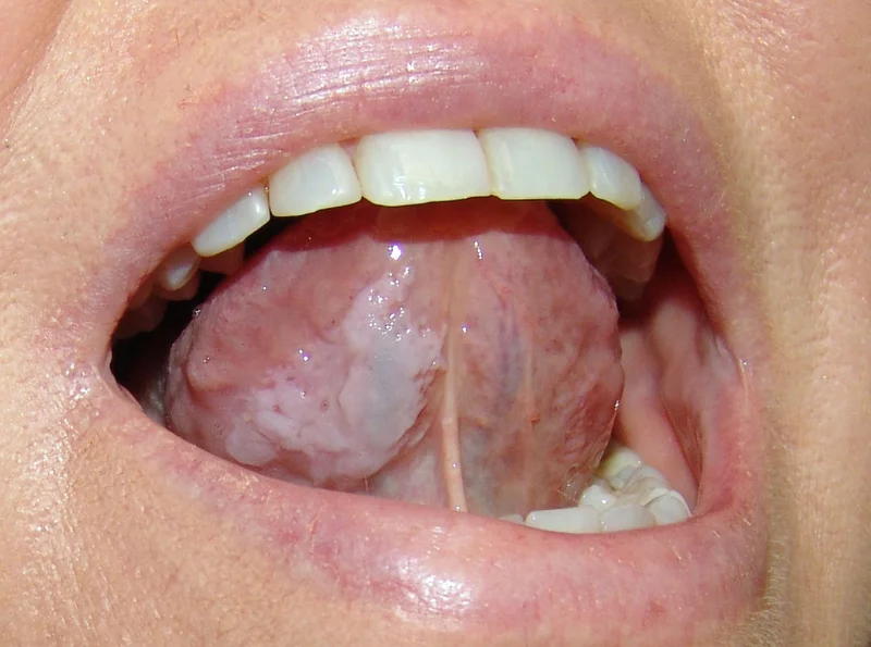
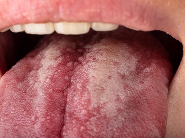
What Does Leukoplakia Look Like?
Leukoplakia presents as a white or grayish-white patch that:
- Cannot be wiped or scraped off (unlike oral thrush, which can be)
- Has been present for at least 2–4 weeks
- May feel slightly raised, thick, or hardened to the touch
- Usually painless, though some patients report mild sensitivity or discomfort, especially with spicy food
Visual Guide by Stage and Severity
| Stage / Severity | Appearance | Texture | Location Tendency | Concern Level |
|---|---|---|---|---|
| Early / Mild | Thin, flat, faintly white patch | Smooth | Buccal mucosa, gums | Low — monitor closely |
| Moderate | Thicker, well-defined white plaque | Slightly rough or leathery | Tongue (lateral border), floor of mouth | Moderate — biopsy recommended |
| Severe / Non-homogeneous | Mixed white-red patch; nodular or verrucous surface | Irregular, bumpy, or corrugated | Floor of mouth, ventral tongue | High — biopsy mandatory |
| PVL (Advanced) | Multifocal verrucous white plaques spreading over time | Wart-like, exophytic | Multiple sites | Very high — aggressive management |
Leukoplakia on Specific Sites
- Leukoplakia on tongue: Most common on the lateral (side) borders. This location carries the highest risk of malignant transformation. Even a small, thin white patch on the side of the tongue warrants evaluation.
- Leukoplakia on gums: Often related to smokeless tobacco placement. Tends to be homogeneous and relatively lower-risk, but still needs monitoring.
- Leukoplakia on lips: Usually the lower lip, sometimes overlapping with actinic cheilitis from sun exposure. Lip lesions have moderate malignant potential.
- Leukoplakia on floor of mouth: High-risk location. Combined with dysplasia, this location has the highest transformation rate.
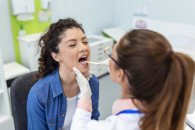
How Is Leukoplakia Diagnosed?
Clinical Examination
- Diagnosis begins with a thorough oral examination. Your dentist or oral medicine specialist will note the size, location, color, texture, and borders of the lesion.
- They will attempt to wipe the patch — if it comes off, it's likely candidiasis, not leukoplakia.
Biopsy: The Gold Standard
A tissue biopsy is essential for any leukoplakia that is non-homogeneous, located in a high-risk area, or persistent.
The biopsy determines:
- Whether dysplasia is present
- The grade of dysplasia: mild, moderate, severe, or carcinoma in situ
- Whether invasive carcinoma has already developed
Histopathological examination evaluates both architectural changes (irregular epithelial stratification, drop-shaped rete ridges, keratin pearls within the epithelium) and cytological changes (nuclear pleomorphism, increased nuclear-to-cytoplasmic ratio, abnormal mitotic figures).
Adjunctive Diagnostic Tools
- Toluidine blue staining: The dye preferentially binds to dysplastic or malignant tissue. Useful as a screening tool, but has limitations with false positives.
- Brush biopsy (OralCDx): A non-invasive technique that collects cells for cytological analysis. Helpful when patients refuse scalpel biopsy, but cannot replace incisional biopsy for definitive diagnosis.
- Autofluorescence (VELscope): Normal mucosa fluoresces green under blue light; dysplastic tissue shows loss of fluorescence. Useful for identifying extent of lesion and guiding biopsy site.
Leukoplakia vs Other White Oral Lesions: Comparison Table
| Condition | Appearance | Can Be Scraped Off? | Risk of Cancer | Key Differentiator |
|---|---|---|---|---|
| Leukoplakia | White/grey plaque, variable texture | No | Yes (3–17.5%) | Diagnosis of exclusion |
| Oral Candidiasis (Thrush) | Creamy white patches | Yes (reveals red base) | No | Positive fungal culture |
| Oral Lichen Planus | White reticular (Wickham's striae) or erosive | No | Low (~1%) | Bilateral, symmetric pattern |
| Leukoedema | Diffuse, milky-white opalescence | No | No | Disappears when mucosa stretched |
| White Sponge Nevus | Thick, white, spongy bilateral patches | No | No | Familial; present from childhood |
| Frictional Keratosis | White, rough patch at site of irritation | No | No | Resolves when irritant removed |
| Oral Hairy Leukoplakia | Corrugated white patches, lateral tongue | No | No | EBV-related; immunosuppressed patients |
This structured comparison is something you won't easily find elsewhere — and it can help you have a more informed conversation with your dentist.
Leukoplakia Treatment: How Is It Managed?
Step 1: Eliminate Risk Factors
The first and most critical step — always — is to stop tobacco use and limit alcohol consumption. In cases caused by chronic irritation, the source (broken tooth, ill-fitting denture) must be corrected. Studies show that approximately 50–60% of leukoplakia lesions regress partially or completely within 3–12 months of tobacco cessation.
Step 2: Observation vs. Intervention
The decision to treat depends on the degree of dysplasia:
- No dysplasia: Active surveillance with clinical examination every 6 months. Re-biopsy if the lesion changes in size, color, or texture.
- Mild dysplasia: Close surveillance every 3–6 months with low threshold for re-biopsy. Some clinicians opt for treatment.
- Moderate to severe dysplasia: Treatment recommended. High risk of progression.
- Carcinoma in situ: Immediate treatment, typically surgical.
Surgical and Procedural Options
| Method | How It Works | Pros | Cons |
|---|---|---|---|
| Conventional surgical excision | Scalpel removal of the lesion | Complete tissue sample for histology | May leave scar, recurrence possible |
| Laser ablation (CO₂ laser) | Vaporizes abnormal tissue | Precise, minimal bleeding, can treat large areas | No tissue specimen for histology |
| Cryotherapy | Freezes lesion with liquid nitrogen | Simple, outpatient, cost-effective | Difficult to control depth; no biopsy specimen |
| Photodynamic therapy (PDT) | Light-activated drug destroys abnormal cells | Minimally invasive, preserves tissue | Limited availability, multiple sessions needed |
| Electrocautery | Burns tissue with electrical current | Widely available | Higher scarring risk |
Recurrence is a major challenge with all methods. A 2018 Cochrane review found no strong evidence that any single treatment approach is superior, and recurrence rates range from 10% to 35% regardless of method.
Nutritional and Chemopreventive Approaches
This is an area that most online resources ignore, but research does exist:
- Vitamin A and retinoids: Several clinical trials, including a landmark study by Hong et al. (1986) in The New England Journal of Medicine, showed that high-dose 13-cis-retinoic acid (isotretinoin) produced regression in 67% of leukoplakia patients. However, toxicity was significant, and relapse occurred after discontinuation in over 50% of cases.
- Beta-carotene: A 1990 study by Stich et al. demonstrated that beta-carotene + vitamin A supplementation led to complete remission of leukoplakia in 27.5% of betel quid chewers vs. 3% in the placebo group.
- Vitamin C: Acts as an antioxidant and may help protect mucosal cells from oxidative damage. Evidence is supportive but not definitive for leukoplakia-specific outcomes.
- Lycopene: A 2007 randomized trial published in Oral Oncology found that lycopene (8mg/day) significantly reduced leukoplakia lesion size compared to placebo.
- Green tea extracts: Preliminary studies suggest epigallocatechin-3-gallate (EGCG) may have antiproliferative effects, but clinical evidence remains limited.
> Important: These nutritional approaches should complement, not replace, medical treatment and follow-up. Discuss supplementation with your healthcare provider before starting.
Can Leukoplakia Be Removed Naturally?
- This is one of the most searched questions online.
- The honest answer: there's no guaranteed natural cure for leukoplakia. However, quitting tobacco, limiting alcohol, maintaining excellent oral hygiene, and optimizing your diet (rich in fruits, vegetables, antioxidants) are natural steps that genuinely help.
- Some lesions — particularly thin, homogeneous ones related to a removable irritant — do resolve on their own once the cause is eliminated.
Be cautious of claims about miracle home remedies. Turmeric, aloe vera, and oil pulling are popular in traditional medicine, but their efficacy for leukoplakia specifically is not supported by robust clinical trials.
Molecular Biomarkers and Predicting Cancer Risk
Understanding which leukoplakia lesions will progress to cancer remains one of the biggest challenges in oral medicine. Beyond histological grading, researchers are investigating molecular biomarkers:
- p53 mutations: The p53 tumor suppressor gene is frequently mutated in dysplastic lesions. Studies show that p53 overexpression in leukoplakia is associated with a 3–4 fold increased risk of malignant transformation.
- Ki-67: A marker of cell proliferation. Elevated Ki-67 index in leukoplakia correlates with higher grades of dysplasia and increased cancer risk.
- Loss of Heterozygosity (LOH): LOH at chromosomes 3p and 9p is considered one of the strongest molecular predictors. A 2012 study in Cancer Prevention Research demonstrated that patients with LOH at these loci had a 22.6-fold increased risk of developing oral cancer.
- DNA ploidy: Aneuploid (abnormal DNA content) leukoplakia lesions have a markedly higher cancer risk compared to diploid lesions.
These biomarkers aren't yet used routinely in clinical practice everywhere, but they are increasingly informing research and may soon become standard in risk stratification — particularly at specialized cancer centers.
Follow-Up Protocol: A Practical Guide
One of the biggest gaps in online information about leukoplakia is a clear, actionable follow-up plan. Here's a practical protocol based on current guidelines and expert consensus:
| Clinical Scenario | Recommended Follow-Up | When to Re-Biopsy | When to Seek Urgent Care |
|---|---|---|---|
| Homogeneous, no dysplasia | Every 6–12 months | If lesion changes size, texture, or color | New pain, bleeding, induration, rapid growth |
| Homogeneous, mild dysplasia | Every 3–6 months | Every 12–18 months, or sooner if change observed | Any red component appears, ulceration develops |
| Non-homogeneous or moderate dysplasia | Every 3 months | Every 6–12 months | Any change in lesion character |
| Severe dysplasia / CIS (treated) | Monthly for first 3 months, then every 3 months for 2 years, then every 6 months | At any sign of recurrence | Any new lesion at or near the treated site |
| PVL | Every 2–3 months indefinitely | Annually or at any change | New lesions, verrucous changes, pain |
Key principle: Leukoplakia patients require lifelong surveillance. Even after successful treatment, recurrence can happen years later. Don't skip appointments.
The Psychological Impact of a Leukoplakia Diagnosis
Something rarely discussed: receiving a diagnosis of a "potentially precancerous" condition causes real anxiety. Patients often catastrophize, assume the worst, or develop health anxiety that affects their daily life.
If you've been diagnosed with leukoplakia, here's what might help:
- Understand the numbers. Most leukoplakia does not become cancer. Even in higher-risk subtypes, regular monitoring catches changes early.
- Stay informed but don't doom-scroll. Stick to reputable sources (peer-reviewed articles, major medical centers). Avoid forums filled with worst-case scenarios.
- Communicate with your doctor. Ask specific questions: "What is my individual risk level? What changes should I watch for? How often should I come back?"
- Consider counseling. If anxiety about the diagnosis is affecting your sleep, appetite, or ability to function, speaking with a mental health professional is completely reasonable and nothing to be ashamed of.
Frequently Asked Questions (FAQ)
Will leukoplakia go away on its own?
- It depends.
- Leukoplakia caused by a clear irritant — like tobacco use or a rough tooth edge — may resolve after the irritant is removed. Studies suggest that 50–60% of tobacco-related lesions improve significantly within a year of quitting. However, idiopathic leukoplakia (without a known cause) is less likely to resolve spontaneously and requires closer monitoring.
Does leukoplakia always become oral cancer?
- No. The overall malignant transformation rate is estimated at 3–17.5%, depending on the study population and follow-up duration. Most leukoplakia lesions remain benign, especially homogeneous ones without dysplasia.
- However, certain subtypes — particularly PVL and non-homogeneous lesions with severe dysplasia — carry a much higher risk.
How much does leukoplakia treatment cost?
Costs vary significantly by country, treatment method, and healthcare system. In India, a scalpel biopsy may cost ₹1,500–₹5,000, while laser ablation can range from ₹5,000–₹25,000 per session depending on the hospital. In the US, costs are substantially higher, often $500–$3,000+ for excisional procedures, and insurance coverage varies. Most health insurance plans in India cover biopsy and surgical treatment when medically indicated.
What questions should I ask my doctor?
- What type of leukoplakia do I have?
- Did the biopsy show any dysplasia? If so, what grade?
- What is my individual risk of this becoming cancer?
- Do I need treatment, or is monitoring enough?
- How often should I come for follow-up?
- Are there lifestyle changes that will help?
- When should I come back sooner than scheduled?
Is leukoplakia contagious?
No. Leukoplakia is not an infection and cannot be transmitted from person to person. Oral hairy leukoplakia is associated with EBV, which is transmissible, but the condition itself develops only under specific immunocompromised conditions.
Conclusion: Take It Seriously, But Don't Panic
Leukoplakia is a condition that deserves your attention — but not your terror. The vast majority of cases are manageable with appropriate lifestyle changes, regular monitoring, and timely intervention when needed. The single most impactful thing you can do is quit tobacco in all forms. Beyond that, maintain regular dental visits, don't ignore changes in your mouth, and if a white patch persists for more than two weeks, get it evaluated.
If you've already been diagnosed, stay consistent with your follow-up schedule and maintain open communication with your healthcare team. Early detection of dysplastic changes transforms a potentially dangerous situation into a highly treatable one.
- Your next step: Schedule an appointment with a dentist or oral medicine specialist if you have any persistent white patch in your mouth. If you're already being monitored, make sure your next follow-up is on the calendar.
- Don't delay — your future self will thank you.
Scientific Sources
- Interventions for treating oral leukoplakia to prevent oral cancer — Lodi G et al., 2016, The Cochrane database of systematic reviews
- Leukoplakia and Immunology: New Chemoprevention Landscapes? — Grigolato R et al., 2020, International journal of molecular sciences
- Traditional Chinese medicine and oral diseases: today and tomorrow — Zheng LW et al., 2011, Oral diseases
- The White Lesion, Hyperkeratosis, and Dysplasia — Ali SA et al., 2019, Otolaryngologic clinics of North America
- Proliferative verrucous leukoplakia: An update — Munde A et al., 2016, Journal of cancer research and therapeutics
- Sanguinaria canadensis: Traditional Medicine, Phytochemical Composition, Biological Activities and Current Uses — Croaker A et al., 2016, International journal of molecular sciences
- Areca nut: a review — Arjungi KN, 1976, Arzneimittel-Forschung
- Chemoprevention of oral cancer: A review and future perspectives — Liao YH et al., 2023, Head & neck
- Pathology-inspired collagen-binding thermosensitive micelle drops enable prolonged and efficient treatment of fungal keratitis — Cai M et al., 2025, Bioactive materials
- Research progress on the role of decorin in the development of oral mucosal carcinogenesis — Rao Y et al., 2025, Oncology research
- Early detection in oral cancer: are we ready for AI-driven precision? — Kumari S et al., 2026, Annals of medicine and surgery (2012)
- The Therapeutic Effects of Curcumin on Oral Disease: A Systematic Review — Indriyani N et al., 2025, Clinical pharmacology : advances and applications
- Anti-inflammatory and Antioxidant Activity of Cucumis sativus and Citrus macroptera Herbal Formulation: An In-Vitro Study — Amani T et al., 2024, Cureus
- Biomarkers for Malignant Potential in Vocal Fold Leukoplakia: A State of the Art Review — Wan P et al., 2021, Otolaryngology--head and neck surgery : official journal of American Academy of Otolaryngology-Head and Neck Surgery
- Chinese Chronic Mucocutaneous Candidiasis: A Case Report Series — Qian G et al., 2024, Infection and drug resistance
- Huli oral health — Newell PL, 2002, Papua and New Guinea medical journal
- Photodynamic therapy (PDT) for oral leukoplakia: a systematic review and meta-analysis of single-arm studies examining efficacy and subgroup analyses — Zhang R et al., 2023, BMC oral health
- DNA integrity in diagnosis of premalignant lesions — Azab NA et al., 2021, Medicina oral, patologia oral y cirugia bucal
- 5-ALA-photodynamic therapy in refractory vulvar lichen sclerosus et atrophicus — Zhang F et al., 2020, International journal of clinical and experimental pathology
- Topical agents for oral cancer chemoprevention: A systematic review of the literature — Chau L et al., 2017, Oral oncology
आयुर्वेदिक डॉक्टर से एक प्रश्न पूछें और मुफ़्त या सशुल्क मोड में अपनी चिंता की समस्या पर ऑनलाइन परामर्श प्राप्त करें।
2,000 से अधिक अनुभवी डॉक्टर हमारी साइट पर काम करते हैं और आपके प्रश्नों की प्रतीक्षा करते हैं और प्रतिदिन उपयोगकर्ताओं को उनकी स्वास्थ्य समस्याओं को हल करने में मदद करते हैं।
संबंधित आलेख


