आयुर्वेदिक डॉक्टर से प्रश्न पूछें और निःशुल्क या भुगतान मोड में अपनी चिंता की समस्या पर ऑनलाइन परामर्श प्राप्त करें। 2,000 से अधिक अनुभवी डॉक्टर हमारी साइट पर काम करते हैं और आपके प्रश्नों का इंतजार करते हैं और उपयोगकर्ताओं को उनकी स्वास्थ्य समस्याओं को हल करने में प्रतिदिन मदद करते हैं।
मुफ्त! आयुर्वेदिक डॉक्टरों से पूछें 24/7
Body DetoxEar, Nose, and Throat DisordersGeriatrics & RejuvenationImmunodeficiencyInfertility TreatmentNutritionOrthopedic DisordersPediatricsRespiratory DisordersSexually Transmitted Diseases (STDs)Surgery RecoveryUrological DisordersVascular DisordersDental DisordersEndocrinological DisordersGastrointestinal DisordersYoga TherapyGeneral MedicineAllergic DisordersCardio DisordersGynecology and ObstetricsCosmetologySkin and Hair DisordersEye DisordersInfectious DiseasesMental DisordersNeurological DisordersOncologyPanchakarmaSexual Health & DisordersSports Medicine
पर प्रकाशित 01/02/25
(को अपडेट 07/02/26)
2,868
Coccydynia Treatment in Ayurveda: Remedies for Tailbone Pain Relief
- Coccydynia is pain centered on the coccyx — the small, triangular bone at the very bottom of your spine, commonly called the tailbone. If you've ever felt a sharp, aching discomfort while sitting down, standing up, or even during bowel movements, you may be dealing with this condition. The good news? Around 90% of coccydynia cases resolve with conservative, non-surgical treatment.
- But understanding the full picture — from anatomy to red flags to specific exercises — is what separates quick recovery from months of unnecessary suffering.
This comprehensive guide covers everything you need to know about coccydynia: what causes it, how its diagnosed, the most effective treatments (including Ayurvedic approaches), and when you should be genuinely concerned. We've structured it to fill the gaps that most medical resources leave open, including differential diagnosis tables, concrete exercise descriptions, and a dedicated section on coccydynia during pregnancy.
What Is Coccydynia?
Definition and Terminology
Coccydynia (pronounced kok-sih-DIN-ee-uh) is the medical term for pain in and around the coccyx. You'll also see it spelled as coccygodynia, or occasionally referred to as coccalgia — all three terms describe the same condition. In plain language, it's simply "tailbone pain."
The term was first introduced by Scottish physician J.Y. Simpson in 1859, though descriptions of coccygeal pain in medical literature date back to the 16th century. Despite its long history, coccydynia remains under-diagnosed and under-treated, partly because many patients feel embarassed discussing pain in this area, and partly because some clinicians dismiss it as trivial.
How Common Is Coccydynia?
Coccydynia accounts for roughly 1% of all spinal pain complaints. It is approximately five times more common in women than in men, primarily due to:
- The wider female pelvis, which leaves the coccyx more exposed
- Childbirth-related trauma
- Greater pressure on the coccyx during seated posture in women
The condition most frequently affects adults, with peak incidence in the fourth decade of life (30–40 years). Adolescents and the elderly are less commonly affected, though it can occur at any age. People who are obese (BMI > 27.4 in women, > 29.4 in men) have increased risk, as do those who have experienced rapid weight loss — the loss of gluteal fat padding removes a natural cushion that protects the coccyx during sitting.
Anatomy of the Coccyx
Understanding coccygeal anatomy helps explain why this small bone can cause such significant pain.
Structure and Variations
The coccyx consists of 3 to 5 fused or semi-fused vertebral segments (Co1–Co5). It articulates with the sacrum above through the sacrococcygeal joint, which normally allows a small degree of flexion and extension — approximately 5° to 25°, with a normal average around 13° of movement during sitting (as measured on dynamic lateral radiographs, per the classification by Maigne, Guedj & Straus).
Key anatomical variations include:
| Morphological Type | Description | Clinical Relevance |
|---|---|---|
| Type I | Coccyx curves gently forward | Normal, lowest pain association |
| Type II | Coccyx curves sharply forward | Moderate risk of coccydynia |
| Type III | Coccyx has a sharp angle/spur | Higher association with pain |
| Type IV | Subluxation at sacrococcygeal or intercoccygeal joint | Strongly associated with coccydynia |
These morphological variants, first described by Maigne et al. in their landmark imaging study, help clinicians identify structural predisposition to coccydynia.
Functions and Attachments
Despite being a vestigial structure, the coccyx serves several important functions:
- Weight-bearing anchor: It forms part of the tripod (along with both ischial tuberosities) that supports body weight when sitting, especially when leaning backward
- Muscle attachment site: The gluteus maximus, levator ani (pelvic floor), and coccygeus muscles all attach to the coccyx
- Ligament anchor: The sacrococcygeal ligament, sacrospinous ligament, and the filum terminale (coccygeal ligament — the terminal end of the spinal cord coverings) all connect here
- Pelvic floor support: Through the levator ani and iliococcygeus, the coccyx is integral to pelvic organ support
This web of attachments explains why coccyx pain can radiate to the hips, lower back, and perineum — and why pelvic floor dysfunction often accompanies chronic coccydynia.
What Causes Coccydynia?
Traumatic Causes
The most common etiology is direct trauma to the coccyx:
- Falls: Landing on the buttocks (the single most frequent cause)
- Childbirth: Vaginal delivery, especially with instrument-assisted birth, can fracture or dislocate the coccyx. Studies suggest coccygeal injury occurs in up to 10% of deliveries
- Repetitive microtrauma: Prolonged cycling, rowing, or sitting on hard, narrow surfaces
- Direct impact: Contact sports injuries
Non-Traumatic Causes
In roughly one-third of cases, no specific trauma is identified.
Non-traumatic causes include:
- Degenerative joint disease of the sacrococcygeal or intercoccygeal joints
- Hypermobility or hypomobility of the coccyx
- Infections: Osteomyelitis, pilonidal cysts with secondary involvement
- Morphological variants: Type III and IV coccyx configurations (described above)
- Referred pain: From lumbar disc herniation, sacroiliac joint dysfunction, or pelvic pathology
- Rapid weight loss: Loss of the ischial fat pad
- Obesity: Increased mechanical loading
What Causes Tailbone Pain Without Injury?
This is one of the most commonly searched questions, and for good reason — many patients are puzzled by coccyx pain that appears "out of nowhere." The answer usually involves one or more of these mechanisms:
- Repetitive low-grade stress from prolonged sitting (especially on hard surfaces) that doesn't register as a single "injury"
- Pelvic floor muscle dysfunction — chronic spasm of the levator ani can create coccygeal pain without any osseous pathology
- Psychogenic factors — depression, anxiety, and somatization disorders have been associated with chronic coccydynia. A 2004 study in the Ochsner Journal noted that patients with "idiopathic" coccydynia had higher rates of psychological comorbidity
- Occult neoplasm — rarely, painless-onset coccygeal pain is the first sign of a chordoma, giant cell tumor, or metastatic disease (from prostate, colorectal, or ovarian cancer)
> Red flag: Coccyx pain without trauma, especially if accompanied by unexplained weight loss, night pain, neurological deficits, or a palpable mass, warrants urgent investigation to rule out malignancy.
What Are the Symptoms of Coccydynia?
Primary Symptoms
The hallmark symptom is localized pain at the coccyx that is:
- Dull and achy at rest, sharp on certain movements
- Worse when sitting, especially on hard surfaces or when leaning backward
- Aggravated by sit-to-stand transitions
- Tender to direct palpation over the coccyx
Associated Symptoms
Depending on severity and cause, patients may also experience:
- Pain during bowel movements or straining
- Pain during sexual intercourse (dyspareunia)
- Aching in the lower back, buttocks, or hips (due to compensatory posture changes)
- Disturbed sleep if rolling onto the back
- Pain during menstruation (in some women)
Impact on Quality of Life
Chronic coccydynia can be surprisingly debilitating. Studies using the Visual Analog Scale (VAS) and Oswestry Disability Index (ODI) show that patients with persistent coccydynia report significant reductions in sitting tolerance, work productivity, and recreational participation. Some patients report being unable to sit for more than 10–15 minutes, effectively limiting driving, desk work, and social activities. In severe cases, coccydynia has been considered grounds for temporary disability classification.
How Is Coccydynia Diagnosed?
Clinical Assessment
Diagnosis begins with a thorough history and physical examination:
- History: Onset, duration, aggravating factors, trauma history, obstetric history, weight changes, bowel/bladder function, and screening for red flags
- Palpation: External and, when indicated, digital rectal examination to assess coccygeal mobility and localize tenderness. The coccyx is grasped between thumb (externally) and index finger (rectally) to check for abnormal mobility, crepitus, or mass
- Provocative maneuvers: Pain reproduced by direct pressure over the coccyx is highly suggestive of coccydynia
What Tests Help Diagnose Coccydynia?
| Test | What It Shows | When It's Used |
|---|---|---|
| Standard lateral X-ray | Fractures, dislocations, spurs | First-line imaging |
| Dynamic (sit/stand) X-ray | Abnormal coccygeal movement (hypermobility or subluxation) | Gold standard for functional assessment |
| MRI | Soft tissue pathology, tumors, infections, bone marrow edema | Suspected neoplasm, infection, or nerve involvement |
| CT scan | Detailed bony anatomy, fracture characterization | Complex fractures, pre-surgical planning |
| Bone scintigraphy | Occult fractures, metastases, infection | Unexplained persistent pain, suspected malignancy |
| Blood tests (ESR, CRP, CBC) | Infection, inflammatory conditions, malignancy screening | Systemic symptoms present |
The dynamic lateral radiograph deserves special mention. By comparing coccygeal position in sitting versus standing, clinicians can measure coccygeal flexion. Movement greater than 25° suggests hypermobility; less than 5° suggests rigidity — both associated with pain.
Differential Diagnosis of Coccyx-Area Pain
- No major medical resource provides a clear, structured differential diagnosis table for coccydynia — which can lead to misdiagnosis.
- Here's a practical comparison:
| Condition | Key Distinguishing Features | How to Differentiate |
|---|---|---|
| Coccydynia | Pain directly over coccyx, tender on palpation, worse with sitting | Positive coccygeal provocation test |
| Pilonidal cyst/abscess | Swelling, redness, drainage in natal cleft | Visual inspection, possible ultrasound |
| Proctalgia fugax | Sudden, severe, brief rectal spasm (seconds to minutes) | Episodic, no coccygeal tenderness |
| Levator ani syndrome | Dull ache in rectum, pressure sensation, lasts >20 min | Tenderness on palpation of levator ani (not coccyx) |
| Piriformis syndrome | Buttock pain radiating down the leg, worse with hip rotation | Positive FAIR test, no direct coccygeal tenderness |
| Perianal abscess | Throbbing perianal pain, fever, swelling | Inspection, systemic signs of infection |
| Sacroiliac joint dysfunction | Pain over SI joint, positive provocative SI tests | Pain localized to SI joint, not midline coccyx |
| Lumbar radiculopathy/sciatica | Radiating leg pain, possible numbness/weakness | Neurological examination, MRI of lumbar spine |
| Chordoma/sacrococcygeal tumor | Progressive pain, palpable mass, neurological deficits | MRI, biopsy |
स्वयं दवा न लें और प्रतीक्षा न करें। अभी डॉक्टर से चैट शुरू करें
How Is Coccydynia Treated?
The treatment of coccydynia follows a stepwise approach. Approximately 90% of patients respond to conservative management, and surgery is reserved for truly refractory cases.
Conservative Treatment (First-Line)
Medications and Protocols
| Medication | Typical Protocol | Notes |
|---|---|---|
| Oral NSAIDs (Ibuprofen, Naproxen) | Ibuprofen 400–600 mg TID or Naproxen 250–500 mg BID for 2–4 weeks | First-line pharmacotherapy; take with food |
| Topical NSAIDs (Diclofenac gel) | Apply to coccygeal area 3–4 times daily | Lower systemic side-effect risk |
| Acetaminophen/Paracetamol | 500–1000 mg every 6–8 hours (max 4g/day) | Alternative if NSAIDs contraindicated |
| Stool softeners (Docusate) | 100 mg BID as needed | To reduce pain during bowel movements |
| Muscle relaxants (Cyclobenzaprine) | 5–10 mg at bedtime, short-term | For associated pelvic floor muscle spasm |
Lifestyle and Ergonomic Modifications
- Coccydynia cushion (wedge/donut cushion): A wedge-shaped cushion with a cutout at the back offloads the coccyx during sitting. This is often the single most effective non-pharmacological intervention
- Avoid prolonged sitting: Take a 5-minute standing break every 30 minutes
- Optimal chair setup: Seat height allowing thighs parallel to the floor, slight forward tilt of the pelvis, feet flat on the ground
- Hot/cold therapy: Ice packs for 15–20 minutes (acute phase, first 48–72 hours), then alternating with warm compresses
Interventional Treatment (Second-Line)
When conservative measures fail after 6–8 weeks, interventional options include:
- Corticosteroid injection (e.g., Triamcinolone 40 mg + 3 mL local anesthetic) into the sacrococcygeal junction — provides relief in approximately 60% of patients
- Ganglion impar (Walther) block: A nerve block targeting the ganglion impar, which provides sympathetic innervation to the coccygeal region. Particularly effective for burning or neuropathic-type coccyx pain
- Manual manipulation: Intrarectal manipulation of the coccyx combined with massage of the levator ani can restore normal mobility. Studies report success rates of 25–30% in chronic cases
- TENS (Transcutaneous Electrical Nerve Stimulation): Non-invasive, can be used at home, modestly effective for pain management
Coccydynia Exercises: Specific Techniques
- Physical therapy is a cornerstone of treatment, yet most resources fail to describe actual exercises.
- Here are evidence-based techniques:
1. Piriformis Stretch
Lie on your back. Cross the affected side's ankle over the opposite knee. Pull the uncrossed leg toward your chest. Hold for 30 seconds. Repeat 3 times on each side.
2. Pelvic Floor Relaxation (Reverse Kegel)
- Sit or lie comfortably. Instead of contracting the pelvic floor (as in a standard Kegel), consciously relax and "lengthen" the pelvic floor muscles as if gently pushing outward. Hold for 5 seconds, repeat 10 times.
- This helps release chronic levator ani spasm — a common contributor to coccydynia.
3. Cat-Cow Stretch (Coccyx Mobilization)
On hands and knees, slowly arch the back upward (cat), then let the belly drop while lifting the tailbone (cow). Focus on movement at the base of the spine. Perform 10–15 repetitions, twice daily.
4. Child's Pose with Pelvic Tilt
From hands and knees, sit back toward your heels with arms extended forward. Gently tuck the pelvis under, then release. Hold each position for 10 seconds. This creates gentle traction on the sacrococcygeal region.
5. Hip Flexor (Iliopsoas) Stretch
Kneel on one knee in a lunge position. Push hips forward gently until a stretch is felt at the front of the hip. Hold 30 seconds. Tight hip flexors alter pelvic tilt and can increase coccygeal loading.
> Important: Avoid exercises that place direct pressure on the coccyx (e.g., sit-ups, certain rowing machine positions) until pain has significantly resolved.
Surgical Treatment (Last Resort)
- Coccygectomy — partial or total removal of the coccyx — is considered only when all conservative and interventional treatments have failed for a minimum of 6–12 months.
- Success rates vary:
| Procedure | Success Rate | Recovery Time | Risks |
|---|---|---|---|
| Partial coccygectomy | 70–80% | 4–8 weeks | Infection (8–10%), wound breakdown, pelvic floor weakness |
| Total coccygectomy | 80–90% | 8–16 weeks | Higher infection risk, rectal injury (rare), persistent pain in 10–20% |
Post-coccygectomy rehabilitation typically involves:
- Weeks 1–2: Wound care, prone/side-lying positions, no sitting
- Weeks 3–6: Gradual reintroduction of sitting (using cushion), gentle pelvic floor exercises
- Weeks 6–12: Progressive return to normal activities, core strengthening
- Months 3–6: Full activity as tolerated
An emerging option for truly refractory cases is spinal cord stimulation (SCS), where electrodes are placed near the sacral nerve roots to modulate pain signals. Evidence remains limited but promising for patients who are not surgical candidates.
Comparison of Treatment Methods
| Method | Effectiveness | Time to Effect | Risk Level | Approximate Cost (India) |
|---|---|---|---|---|
| NSAID + Cushion | 60–70% | 1–4 weeks | Low | ₹200–1,500 |
| Physical therapy | 50–70% | 4–12 weeks | Very low | ₹500–2,000/session |
| Corticosteroid injection | 60% | Days to 2 weeks | Low-moderate | ₹2,000–8,000 |
| Ganglion impar block | 60–75% | Days | Moderate | ₹5,000–15,000 |
| Manual manipulation | 25–30% | Weeks | Low | ₹1,000–3,000/session |
| Coccygectomy | 80–90% | 2–4 months | Moderate-high | ₹50,000–2,00,000 |
Coccydynia During Pregnancy
Tailbone pain is remarkably common during pregnancy and the postpartum period, yet few resources address it adequately.
Why It Occurs
- Relaxin hormone loosens the sacrococcygeal and intercoccygeal ligaments, creating hypermobility
- Increased body weight and shifted center of gravity place greater load on the coccyx
- Vaginal delivery can bruise, dislocate, or fracture the coccyx (risk increases with prolonged labor, large baby, or forceps delivery)
Safe Treatment Options During Pregnancy
| Safe | Use With Caution | Avoid |
|---|---|---|
| Coccyx cushion | Paracetamol (limited duration) | NSAIDs (especially 3rd trimester) |
| Ice/heat therapy | Gentle manual therapy | Corticosteroid injections (without obstetric clearance) |
| Pelvic floor physiotherapy | Topical analgesics | X-rays (unless essential) |
| Side-lying sleeping position | MRI with gadolinium contrast | |
| Prenatal yoga (modified) |
Postpartum coccydynia usually resolves within 3–6 months as hormonal levels normalize and tissue heals. Persistent pain beyond 6 months should prompt imaging and specialist referral.
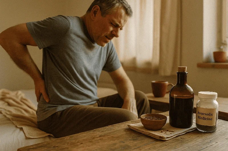
Ayurvedic Perspective on Coccydynia
In Ayurveda, coccydynia is understood primarily as a Vata dosha imbalance affecting the Kati (lower back and pelvic) region. The condition relates to concepts like Katishoola (lower back pain) and Gridhrasi (sciatica-like conditions) when nerve involvement is present.
Ayurvedic Treatment Approaches
- Kati Basti: A dam of dough is placed on the lower back/sacral area and filled with warm medicated oil (typically Mahanarayana taila or Dhanwantharam taila). This provides sustained warmth and herbal absorption for 30–45 minutes
- Abhyanga: Therapeutic oil massage using Bala taila or Ashwagandha taila to pacify Vata and reduce muscle tension around the coccyx
- Pichu: A cotton pad soaked in warm medicated oil placed over the coccygeal area
- Internal medications: Yogaraja Guggulu, Rasnadi Kashayam, and Ashwagandha churna are commonly prescribed to manage pain and inflammation
Integrating Ayurveda with Conventional Treatment
Ayurvedic therapies work best as complementary approaches alongside evidence-based conventional treatment. Many patients in India find significant relief combining NSAID therapy with Kati Basti sessions and specific yoga asanas (modified Bhujangasana, Marjaryasana-Bitilasana). Always inform both your Ayurvedic practitioner and allopathic doctor about all treatments you're receiving.
When to See a Doctor: Red Flag Checklist
Not all tailbone pain is benign.
Seek medical attention promptly if you experience:
- Coccyx pain without any history of trauma or obvious cause
- Unexplained weight loss accompanying the pain
- Night pain that wakes you from sleep
- Fever or chills with coccyx tenderness
- Neurological symptoms: numbness, tingling, weakness in legs, or loss of bowel/bladder control
- Palpable lump or mass near the coccyx
- Pain that is progressively worsening despite 4–6 weeks of conservative treatment
- History of cancer (especially prostate, colorectal, ovarian, or bone cancers)
Does Coccydynia Go Away?
The prognosis for coccydynia is generally favorable. Most cases resolve within 8–12 weeks with conservative treatment. A 2014 review in the Ochsner Journal noted that the majority of patients achieve satisfactory pain relief without surgery. However, a subset of patients (roughly 10%) develop chronic coccydynia lasting more than 3 months, which may require escalation to interventional treatments.
Factors associated with poorer prognosis include:
- Coccygeal hypermobility or subluxation on dynamic X-ray
- Type IV coccyx morphology
- Coexisting psychological conditions (depression, anxiety)
- Obesity
- Delay in treatment initiation
Frequently Asked Questions (FAQ)
Can Coccydynia Be Permanent?
True permanent coccydynia is uncommon. While some patients experience symptoms for years, very few remain permanently symptomatic if treated appropriately. Even refractory cases usually respond to coccygectomy, with 80–90% success rates for total resection.
Is Coccydynia a Disability?
Coccydynia is not typically classified as a permanent disability, but severe cases can qualify for temporary disability benefits if documented inability to sit for occupational requirements is demonstrated. In India, this would need assessment by a medical board. The ICD-10 code for coccydynia is M53.3, which is used for insurance and disability documentation.
How Long Does Coccydynia Last?
- Acute coccydynia (post-trauma): 4–8 weeks with treatment
- Subacute: 8–12 weeks
- Chronic: Beyond 3 months; requires investigation and possibly advanced treatment
Can You Exercise With Coccydynia?
Yes, but selectively. Low-impact activities like swimming, walking, and the specific stretches described above are encouraged. Avoid cycling, rowing, and exercises involving direct coccygeal pressure until pain resolves. Exercise actually aids recovery by improving blood flow and reducing muscle tension.
What Is the Best Pillow for Coccydynia?
A wedge-shaped coccyx cushion with a posterior cutout (U-shaped or V-shaped) is most effective. It redistributes weight to the ischial tuberosities and thighs while offloading the coccyx. Memory foam versions provide the best pressure distribution. Donut cushions work but may cause instability during prolonged sitting.
How Is Coccydynia Seen on Radiology?
Standard lateral X-rays may show fracture, dislocation, or bony spurs. Dynamic (sit/stand) lateral radiographs are the gold standard, revealing abnormal flexion (>25°, suggesting hypermobility) or rigidity (<5°). MRI is used for soft tissue assessment and tumor evaluation.
Conclusion
Coccydynia is a common, treatable condition that responds well to a stepwise approach — starting with simple measures like a coccyx cushion and NSAIDs, progressing to physical therapy, injections, and in rare cases, surgery. The key is early, accurate diagnosis to rule out serious pathology and to match the treatment to the underlying cause.
If you're dealing with persistent tailbone pain, don't dismiss it or wait it out indefinitely. Consult a qualified orthopedic specialist or spine physician for proper evaluation. For those interested in complementary approaches, Ayurvedic therapies like Kati Basti and specific yoga-based exercises can be valuable additions to your treatment plan.
Have questions about your specific case of coccydynia? Our verified Ayurvedic doctors are available 24/7 to provide personalized guidance on managing tailbone pain through holistic, evidence-informed approaches.
Scientific Sources
- Anococcygeal Nerve and Sitting Pain: Differential Diagnosis and Treatment Results — Alimehmeti RH et al., 2022, Annals of plastic surgery
- Minimally invasive exoscope-assisted coccygectomy: A novel approach for chronic refractory coccydynia — Obeng-Gyasi B et al., 2025, World neurosurgery: X
- Advances in Coccygectomy: A Comprehensive Review Evaluating Surgical Techniques for Coccygodynia — Obeng-Gyasi B et al., 2025, Brain sciences
- Laser acupuncture for refractory coccydynia after traumatic coccyx fracture: A case report — Lin CH et al., 2020, Medicine
उपयोगकर्ताओं के प्रश्न
What is the best type of exercise to help relieve coccydynia pain?
Isaac
5 दिनों पहले
For coccydynia pain, try gentle exercises like pelvic tilts, and stretching the lower back and hips. These moves can help in increasing blood flow and reducing tension. Avoid sitting for too long or high-impact exercises. It's always a good idea to listen to your body and not push through pain.
What causes coccydynia and how can I prevent it from happening?
Grayson
14 दिनों पहले
Coccydynia is usually caused by trauma to the coccyx like falls, childbirth, or just prolonged sitting—sometimes it's not obvous why it happens. To prevent it, you can try using cushioned seats, taking breaks from sitting too long, and doing gentle stretches to maintain mobility and strength in the hip area. Also, keep a balanced posture to avoid extra pressure on the coccyx.
What type of physical therapy exercises are most effective for coccydynia relief?
Aria
23 दिनों पहले
For coccydynia, Cat-Cow Stretch is great as it helps in coccyx mobilization. Basically, you're on all fours, arch your back up like a cat, then lower it like a cow, hold each for 10 secs. Ankle-over-knee stretch can also be helpful. Avoid activities putting direct pressure on the tailbone like cycling until the pain eases a bit. Trying a TENS unit might also give some relief at home!
Can I use essential oils to help manage coccydynia symptoms effectively?
Anthony
34 दिनों पहले
Yes, essential oils can be helpful! Oils like lavender or peppermint can offer some relief due to their anti-inflammatory and pain-relieving properties. Just a bit of them mixed with a carrier oil, then massaged into the lower back might help. But remember everyone's different, so it's good to check with your practitioner to tailor it to your dosha balance and any other specifics in your lifestyle.
Is it safe to use Bhringraj oil for pain relief in coccydynia?
Virginia
44 दिनों पहले
Bhringraj oil isn't typically used for pain relief like coccydynia, especially as its more known for hair and skin benefits. However, it's generally safe if manufactured properly. For pain, I'd suggest considering oils with proven anti-inflammatory benefits like Guggulu. It's always good to check with a practitioner for tailored advice!
Can Ashwagandha be used safely with other supplements for coccydynia treatment?
Matthew
54 दिनों पहले
Yes, Ashwagandha can generally be safely combined with other supplements for coccydynia, but it's important to be mindful of how these supplements interact and affect your Vata dosha. Always pick high-quality supplements and keep an eye on the dosages. It's a good idea to talk to a healthcare provider to tailor it to ya needs, just to be safe!
What is Bilwadi Taila and how does it help with pain relief?
Yvonne
63 दिनों पहले
Bilwadi Taila is an ayurvedic herbal oil that's great for soothing muscle pain and inflammation. When you apply it, it helps relax muscles and reduce swelling, making it effective for pain relief, including coccydynia. It's especially good for balancing the Vata dosha, which, when imbalanced, can cause pain and discomfort.
What is the role of Vata dosha in coccydynia and how can it impact treatment?
Mateo
73 दिनों पहले
Vata dosha, when imbalanced, can aggravate coccydynia by causing dryness, pain, and stiffness in the coccyx region. This makes it crucial to focus on grounding and warm therapies during treatment. Herbal oils like ashwagandha and warming foods can help pacify Vata, reduce pain, and enhance mobility.
Definitely consult an ayurvedic practitioner to tailor the treatments!
Can I use meditation or yoga to help manage coccydynia pain effectively?
Maya
82 दिनों पहले
Yes, definitely! Meditation and yoga can be great for managing coccydynia pain. They help balance the Vata dosha, which may be contributing to the pain. Gentle yoga poses like child's pose or meditative breathing can soothe nerves and reduce tension in the tailbone area. Just be sure to listen to your body and avoid anything that causes pain or discomfort. 😌
Is it safe to use Ashwagandha for coccydynia if I have allergies?
Teagan
92 दिनों पहले
Using Ashwagandha could be helpful for coccydynia because of its anti-inflammatory properties, but when you have allergies, it's always best, you know, to proceed carefully. Maybe do a patch test or check with an Ayurvedic practitioner to make sure it won't trigger any allergic reactions. Safety first!
संबंधित आलेख

General Medicine
Kushmanda Rasayana for Weight Gain – Ayurvedic Solution for Healthy Weight Management
Discover how Kushmanda Rasayana can help with weight gain, including its Ayurvedic ingredients, therapeutic benefits, and how it promotes overall health and nourishment for natural weight gain.
4,303

General Medicine
Nagaradi Choornam: Ayurvedic Uses, Benefits & Dosage Guide
Discover the benefits, uses, dosage, and Ayurvedic insights of Nagaradi Choornam. Learn how this traditional remedy supports digestive health and vitality.
3,886

General Medicine
Haridra Khanda for Cold – Ayurvedic Herbal Remedy for Respiratory Relief
Discover Haridra Khanda, an Ayurvedic herbal formulation renowned for relieving cold symptoms. Learn how Haridra Khanda balances doshas, reduces inflammation, and supports respiratory health naturally
3,953

General Medicine
Perment Capsules Uses: Benefits, Dosage & Scientific Insights
Discover the uses of Perment Capsules. Learn about their benefits, proper dosage, usage guidelines, and scientific insights to enhance your well-being naturally.
4,309

General Medicine
Gruhadhoomadi Choornam: Ayurvedic Formula for Digestive Wellness
Learn about Gruhadhoomadi Choornam, a potent Ayurvedic herbal powder that supports healthy digestion and overall vitality. Discover its traditional benefits for balancing your body.
2,276

General Medicine
एट्रिसोर कैप्सूल
एट्रिसोर कैप्सूल की खोज
1,237

General Medicine
Lohasava Uses, Dose, Side Effects, And Ingredients
Exploration of Lohasava Uses, Dose, Side Effects, And Ingredients
2,313
 - Ask Ayurveda Image")
General Medicine
Ayurvedic Medicine for Body Pain and Weakness: What Actually Works (And Why It's Not Just in Your Head)
Ayurvedic medicine for body pain and weakness isn’t just a pill or a cream. It’s a whole philosophy of how the body functions, breaks down, and rebuilds. It treats fatigue and pain not as isolated issues but as signals — whispers from within that somethin
3,589

General Medicine
What Causes Sleep Apnea?
What causes sleep apnea? Learn about the root causes of central and obstructive sleep apnea, its link to weight gain and high blood pressure
2,023

General Medicine
Sadhaka Pitta: Understanding its Role in Mind-Body Balance
Explore the science behind Sadhaka Pitta, its health implications, and evidence-based insights. Learn how it influences emotional well-being and cognition.
3,100
विषय से संबंधित परामर्श
अतिरिक्त दस्तावेज़
© 2024 Ask Ayurveda. सर्वाधिकार सुरक्षित।